Loculated Pleural Effusion Cxr ~ Dark lung fields. Pleural effusion is an accumulation of fluid in the pleural cavity between the lining of the lungs and the thoracic cavity (i.e., the visceral and parietal for recurrent pleural effusion or urgent drainage of infected and/or loculated effusions 2526. Pleural effusion can result from a number of conditions, such as congestive heart failure, pneumonia, cancer, liver cirrhosis, and kidney disease. Pleural fluid/serum ldh ratio >0.6. Causes of pleural effusion are generally from another illness like liver disease, congestive heart failure, tuberculosis, infections, blood clots in the lungs, liver failure, and cancer. Effusion on cxr—> free fluid (not loculated)—> fluid >1cc—> next step.
Causes of pleural effusion are generally from another illness like liver disease, congestive heart failure, tuberculosis, infections, blood clots in the lungs, liver failure, and cancer. Differentiation of loculated effusions from solid masses. This situation most commonly is seen in patients with heart failure. Detection of pleural effusion(s) and creation of initial differential diagnosis are a pleural effusion of 500 ml will obscure diaphragmatic contour on upright cxr; Large pleural effusions, s/p thoracentesis with pleural fluid suggestive of transudative process.
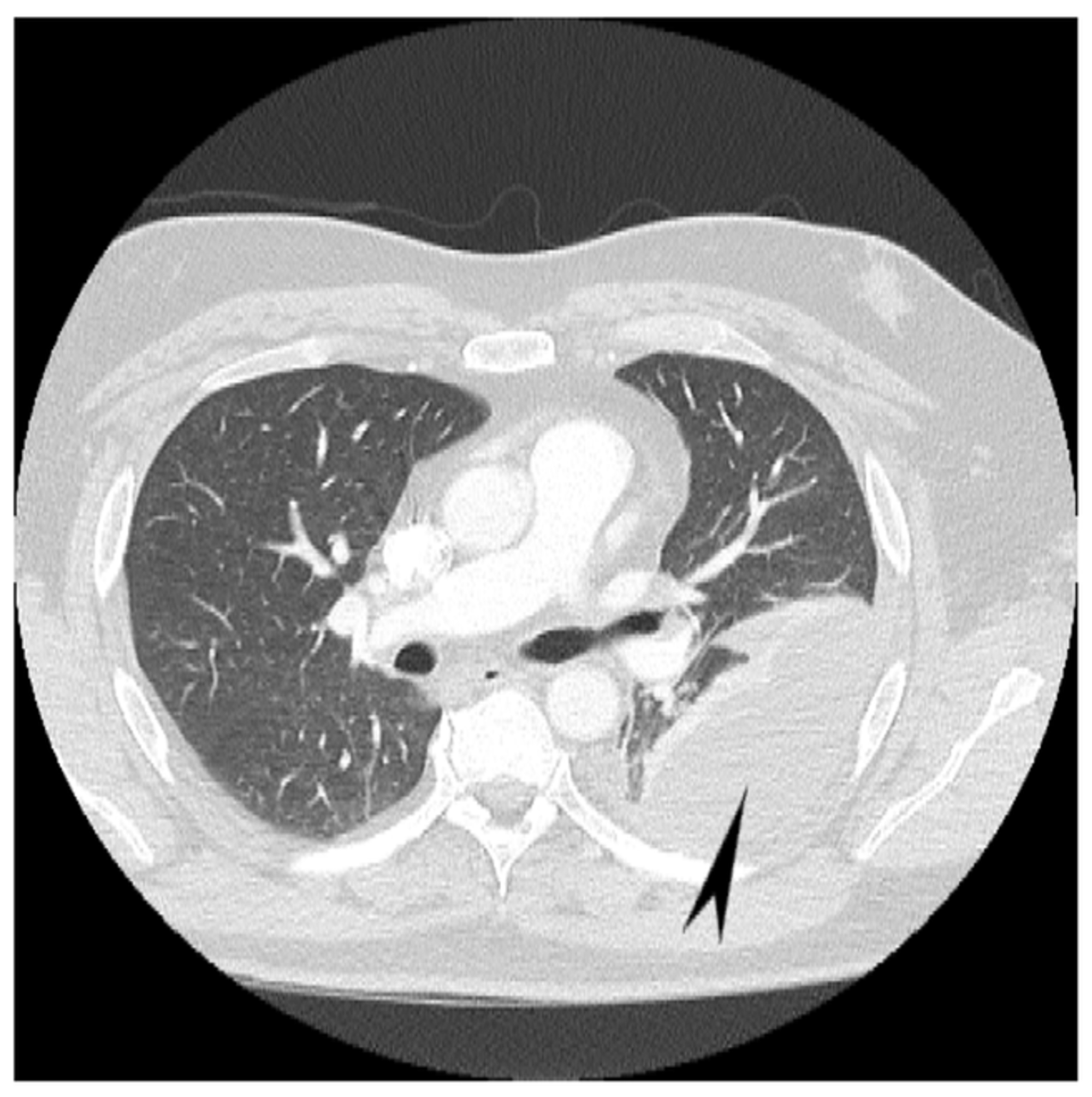
Preoperative CXR showing the loculated empyema. | Download Scientific Diagram from www.researchgate.net The lungs and the chest cavity both have a lining that consists of pleura, which is a thin membrane. Pleural fluid/serum ldh ratio >0.6. Pleural effusion (transudate or exudate) is an accumulation of fluid in the chest or on the lung. Pleural effusion, popularly known as water in the pleura or water in the lung, is the name given to the abnormal accumulation of fluid in the pleura, a thin membrane surrounding the lung. Case contributed by dr prashant mudgal. Treatment depends on the cause. Dr bhatia discussing on pleural effusion in #lastminuterevisionpointdiscussionseries. Pleural effusions occur as a result of increased fluid formation and/or reduced fluid resorption.
Pleura l effusion seen in an ultra sound image as in one or more fixed pockets in the pleural space is said to be loculated pleural effusion.in.
Treatment depends on the cause. Loculated pleural effusion on cxr. Approximately 1 million people develop this abnormality each year in the united states. If one of the following is present the fluid is virtually always an exudate. Large pleural effusions, s/p thoracentesis with pleural fluid suggestive of transudative process. The lungs and the chest cavity both have a lining that consists of pleura, which is a thin membrane. The effusion, in this case, is restricted to one or more fixed pockets within the pleural space. 9 633 просмотра 9,6 тыс. Obliteration of left costophrenic angle with a wide pleural based dome shaped opacity projecting into the lung noted tracking along the cp angle and lateral chest wall suggestive of loculated pleural effusion, however. Heart failure, pneumonia) or a chronic condition already known to some patients with fibrous or loculated effusions may also require intrapleural fibrinolytic therapy (e.g. Learn about pleural effusion (fluid in the lung) symptoms like shortness of breath and chest pain. It detects pleural effusions with higher sensitivity and specificity than cxr, and provides valuable information about the size and depth of the pleural effusion, the echogenicity of the fluid, the presence of septated or loculated fluid, pleural thickening and nodularity, and the presence of any. Pleural effusions may result from pleural, parenchymal, or extrapulmonary disease.
A pleural effusion is accumulation of excessive fluid in the pleural space, the potential space that surrounds each lung. A pleural effusion may be malignant (caused by cancer) or nonmalignant (caused by a condition that is not cancer). Determine if it can be tapped. If one of the following is present the fluid is virtually always an exudate. Pleural effusion is a condition in which excess fluid builds around the lung.
Pleural effusion | Radiology Reference Article | Radiopaedia.org from prod-images.static.radiopaedia.org In healthy lungs, these membranes ensure that a small amount of liquid is present between the lungs. There is a large left pleural effusion obscuring the lower half of the left hemi thorax. Pleural effusion can be a sign of serious illness. It detects pleural effusions with higher sensitivity and specificity than cxr, and provides valuable information about the size and depth of the pleural effusion, the echogenicity of the fluid, the presence of septated or loculated fluid, pleural thickening and nodularity, and the presence of any. Pleural fluid/serum protein ratio >0.5. 9 633 просмотра 9,6 тыс. Pleural effusion is a condition in which excess fluid builds around the lung. Tx if pt has chf.
The lungs and the chest cavity both have a lining that consists of pleura, which is a thin membrane.
Pleural effusion is not a disease, but a common manifestation of several different diseases. Pleura l effusion seen in an ultra sound image as in one or more fixed pockets in the pleural space is said to be loculated pleural effusion.in. e intrinsic characteristics of an effusion and its. Large right effusion (red arrow) displacesthe heart to the left (yellow arrow). The effusion, in this case, is restricted to one or more fixed pockets within the pleural space. Pleural fluid/serum protein ratio >0.5. Pleural fluid ldh > two thirds of upper limit for serum ldh. Bhatia medical coaching institute, dbmci. Pleural effusion occurs when too much fluid collects in the pleural space (the space between the two layers of the pleura). Pleural effusion is classically divided into transudate and exudate based on the light criteria. Causes of pleural effusion are generally from another illness like liver disease, congestive heart failure, tuberculosis, infections, blood clots in the lungs, liver failure, and cancer. It detects pleural effusions with higher sensitivity and specificity than cxr, and provides valuable information about the size and depth of the pleural effusion, the echogenicity of the fluid, the presence of septated or loculated fluid, pleural thickening and nodularity, and the presence of any. Accompanying adhesions can be identified.
Pleural fluid ldh > two thirds of upper limit for serum ldh. Approximately 1 million people develop this abnormality each year in the united states. Pleural effusion, popularly known as water in the pleura or water in the lung, is the name given to the abnormal accumulation of fluid in the pleura, a thin membrane surrounding the lung. no change in position of effusion withchange in position of chest. Pleural effusions occur as a result of increased fluid formation and/or reduced fluid resorption.
Cureus | Hemorrhagic Pleural Effusion: A Rare Presentation of Vitamin K Deficiency in an Adult ... from assets.cureus.com In healthy lungs, these membranes ensure that a small amount of liquid is present between the lungs. This situation most commonly is seen in patients with heart failure. e intrinsic characteristics of an effusion and its. What does pleural effusion mean? Accompanying adhesions can be identified. Loculated pleural effusion on cxr. Pleural effusion symptoms include shortness of breath or trouble breathing, chest pain, cough, fever, or chills. Pleural effusion can be a sign of serious illness.
The pleura are thin membranes that line the lungs and the inside of the chest cavity and act to lubricate and facilitate breathing.
Approximately 1 million people develop this abnormality each year in the united states. Treatment depends on the cause. oracentesis of loculated pleural effusions clinical quiz. Meaning of pleural effusion medical term. Pleural fluid/serum protein ratio >0.5. Determine if it can be tapped. A pleural effusion is accumulation of excessive fluid in the pleural space, the potential space that surrounds each lung. Loculated pleural effusion on cxr. Dr bhatia discussing on pleural effusion in #lastminuterevisionpointdiscussionseries. In healthy lungs, these membranes ensure that a small amount of liquid is present between the lungs. There is a large left pleural effusion obscuring the lower half of the left hemi thorax. Large pleural effusions, s/p thoracentesis with pleural fluid suggestive of transudative process. Pleural fluid/serum ldh ratio >0.6.
Treatment depends on the cause loculated pleural effusion. no change in position of effusion withchange in position of chest.
Share :
Post a Comment
for "Loculated Pleural Effusion Cxr ~ Dark lung fields"


Post a Comment for "Loculated Pleural Effusion Cxr ~ Dark lung fields"